Polarized Microscopy: Understanding the basics

Simple brightfield microscopes are great. They can be used to study a wide variety of specimens with relatively simple set up. However, for industrial researchers, or geological sciences, there are some objects that are tricky to view, particularly objects that have a lot of glare or birefringence. This is where polarized microscopy can save the day!
How it works
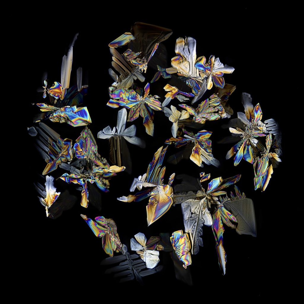
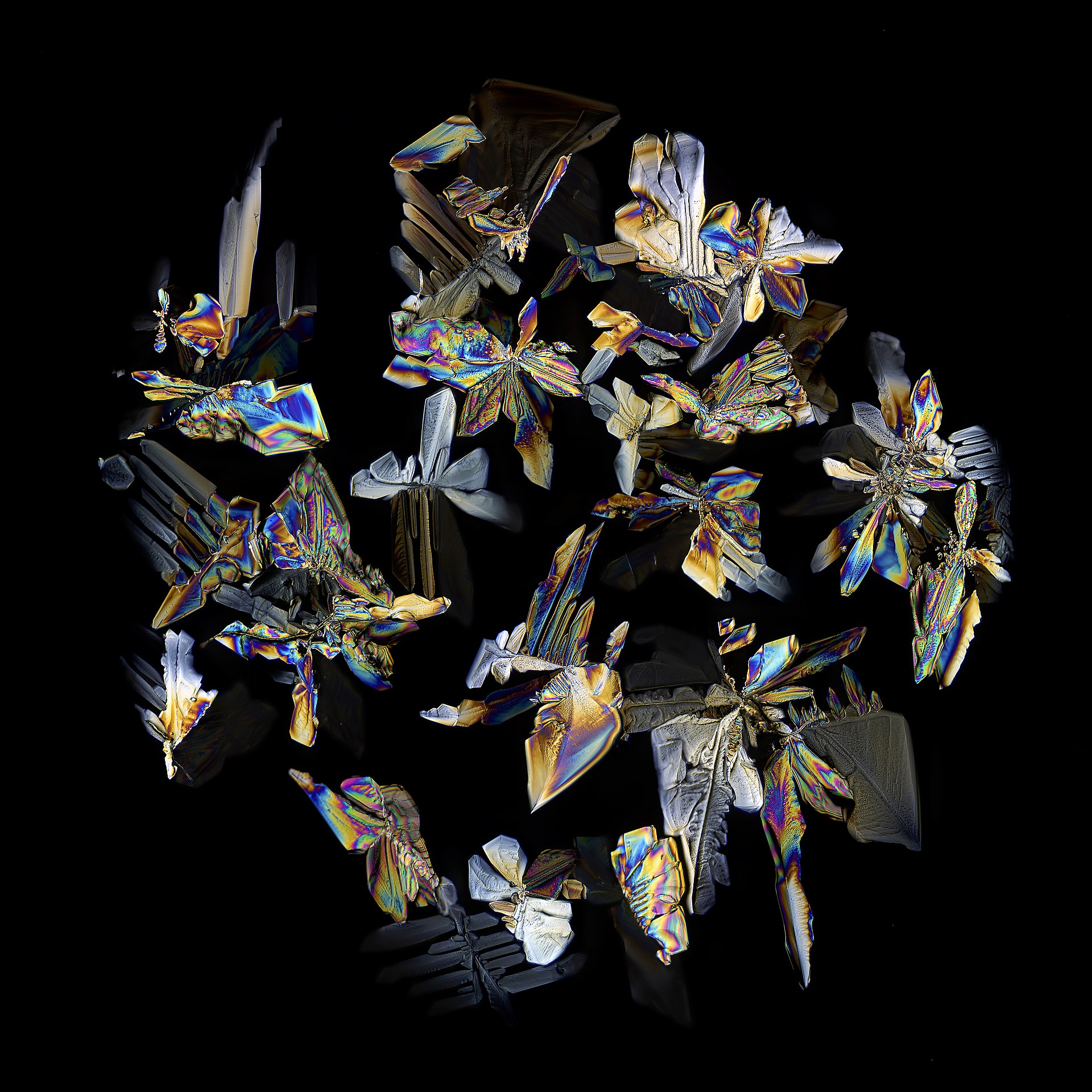
Polarized light microscopy is specifically for the examination of anisotropic (birefringent) objects such as minerals, polymers, crystals, and more. There are plenty of applications in metallurgical fields as well.
These types of microscopes differ from ordinary microscopes by having a precisely centered, round, rotating stage. Furthermore, specialized filters above and below the stage help filter the light to produce a clearer image.
One filter, referred to as the polarizer, is located in the light path beneath the sample slide. Through it the object is illuminated with linear polarized light.
The second filter, called the analyzer, is situated in the observation beam path allowing the analysis of the linear polarized light modified by the object.
When the polarizer and the analyzer are “crossed” (referred to as crossed polars) it means that there is a 90° difference in the vibration plane of the light allowed through them. When no sample is present, or an isotropic material is in the beam path, no light will come through.
As a result, polarized microscopy allows us to see objects with much less reduced glare.
Advantages of polarized microscopy
Polarized microscopy improves the quality of the image of reflective materials, especially when compared to traditional brightfield and darkfield illumination. Highly reflective metal surfaces will have little to no glare allowing us to clearly see the details behind the glare.
Furthermore, polarized light microscopes have a high degree of sensitivity and can provide both quantitative and qualitative data targeted at a wide range of anisotropic specimens.
This field of polarized microscopy, is also quite popular among geologists, mineralogists, and chemists. As a result, there is a lot of documentation and scientific research around its use and application. However, there is less support when it comes to applications in life sciences.
Limitations of polarized microscopy
As stated above, using polarized microscopy in the life sciences has limitations. Biological specimens, such as histological sections, have birefringent materials that are oriented at different angles throughout the sample. This can make imaging and interpretation more difficult. A simple brightfield microscope is sufficient for these applications.
Lastly, while polarized microscopy is quite popular among geologists, mineralogists, and chemists, it requires a lot of training to apply it properly. This means that there is a higher bar of entry of the field and longer training periods. Even so, users will be analyzing samples and diffraction patterns within minutes for routine applications.

